


Meniscus Tear Grades Explained: What Grade 1–4 Really Means
A meniscus tear diagnosis often comes with a grade attached, and for many patients, that number becomes the focus of concern. A “grade 3” tear can sound alarming, while a “grade 1” tear might be dismissed as minor. In reality, the grading system used on MRI reports measures something quite specific, and it does not tell the whole story on its own. Understanding what these grades actually describe, and how they relate to symptoms and treatment, helps make sense of a diagnosis that can otherwise feel confusing.
What Meniscus Grading Actually Measures
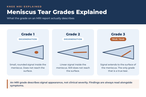
The grading system most commonly referenced (known as the Stoller classification) describes the appearance of signal intensity within the meniscus on MRI, not the severity of a tear, as most patients assume.
Grade 1: A small, rounded area of increased signal within the meniscus that does not reach the surface. This reflects early degenerative change within the cartilage rather than an actual tear.
Grade 2: A linear area of increased signal within the meniscus that does not extend to the articular surface. This also represents degeneration rather than a true tear, though it is more advanced than Grade 1.
Grade 3: Increased signal that extends to at least one surface of the meniscus. This is the only grade that represents an actual tear in the structural sense.
This distinction matters. Grades 1 and 2 are MRI descriptions of degenerative change within meniscal cartilage, a normal part of ageing in many knees, and are not tears at all. Only a Grade 3 finding reflects a genuine tear that has disrupted the meniscus surface.
Beyond the Grade: Tear Pattern and Location
For a true (Grade 3) tear, the grade alone does not capture the full clinical picture. Other factors carry significant weight in determining management, including:
Location: Tears in the outer third of the meniscus (the “red zone”) have a blood supply and greater capacity to heal. Tears in the inner two-thirds (the “white zone”) have little to no blood supply, limiting healing potential.
Pattern: Whether the tear is a simple horizontal or vertical split, a radial tear, or a more complex bucket-handle or root tear affects both symptoms and available surgical options.
Stability: Some tears are stable and unlikely to progress or cause mechanical symptoms, while others cause the torn fragment to catch or lock within the joint.
Key takeaway: An MRI grade describes signal appearance, not clinical severity. A Grade 3 tear in the well-vascularised outer meniscus with a simple pattern often behaves very differently to a Grade 3 tear that is complex, unstable, and located in the poorly vascularised inner zone.
Why Imaging Must Be Read Alongside Symptoms
It is well recognised that meniscal degeneration visible on MRI, including Grade 3 signal change, is extremely common in people over 40 with no knee symptoms at all. This means a scan cannot be interpreted in isolation.
The clinical picture, including where the pain is felt, whether there is swelling, catching, locking, or giving way, and how the knee responds to movement and loading, is essential to understanding whether an MRI finding is actually the source of a patient’s symptoms or an incidental finding.
Clinical note: A high MRI grade does not automatically mean surgery is required, and a low grade does not rule out a clinically significant tear. Persistent mechanical symptoms such as locking or a knee that gives way warrant assessment regardless of the reported grade.
How Grade and Pattern Influence Management
Once a tear has been assessed in the context of symptoms, management generally follows a structured approach:
- Modify painful activities. Reducing movements that provoke mechanical symptoms, such as deep squatting, pivoting, or twisting, allows irritated tissue to settle without requiring complete rest.
- Control inflammation. Addressing joint swelling and irritation supports comfort and function while the underlying tear pattern and its behaviour become clearer over time.
- Progressive strengthening. Building strength around the knee, particularly through the quadriceps and hip musculature, supports the joint and can reduce load through the injured portion of the meniscus.
Degenerative changes (Grades 1 and 2) rarely require anything beyond this kind of conservative management. True tears (Grade 3) that are stable, small, and located in the well-vascularised outer zone are also frequently managed this way with good outcomes. Tears that are unstable, causing persistent mechanical symptoms, or located in a zone with limited healing capacity are more likely to be discussed in terms of arthroscopic options.
Getting the Full Picture
A meniscus tear grade is useful, but it is only one part of a larger clinical assessment. Understanding tear location, pattern, and how the findings correlate with a patient’s actual symptoms allows for a management plan tailored to the individual knee rather than the number on a report.
Anyone with persistent knee pain, swelling, or mechanical symptoms following a suspected meniscus injury should have their imaging reviewed alongside a thorough clinical examination to determine the most appropriate path forward.
When to Seek Assessment
If knee pain, swelling, or mechanical symptoms such as catching or locking persist or affect daily activity, a formal assessment can clarify what an MRI grade actually means for that specific knee. Dr Daniel Cohen consults at Bondi Junction and Kogarah, offering thorough clinical assessment alongside imaging review to guide an appropriate, individualised management plan.